When we think about how living things grow and repair themselves, it's pretty wild to consider the tiny, invisible actions happening inside every single cell. It's almost like a carefully choreographed dance, a sequence of events that has to unfold just so, you know, for everything to work out right. We often talk about these distinct periods, these specific moments in time where something changes, as "phases." Just like the moon goes through its regular changes in how it looks, or how a project moves from one particular period to the next, cells also have their own set of steps they follow.
For something as fundamental as cell division, which is what mitosis is all about, these distinct periods are really what help us make sense of the whole thing. It’s a process where one cell becomes two, identical copies, and this isn't just a sudden snap of the fingers kind of deal. No, it actually happens in a series of very clear, observable steps, each one building on the one before it. Thinking about it, it's a lot like how a story unfolds, with different chapters that each move the plot along, you see.
So, when we put these steps up on a whiteboard, it really helps to visualize how a cell changes its appearance and what it’s doing at each particular moment. It helps us to grasp how a cell goes from being one thing to becoming two, with all the bits and pieces inside moving around and getting ready for the big split. This way of looking at the phases of mitosis on the whiteboard makes a rather complex idea much more approachable, you know, like breaking a big task into smaller, easier-to-manage parts.
Table of Contents
- What Are the Phases of Mitosis, Anyway?
- Prophase on the Whiteboard - How Does It Start?
- Metaphase - Lining Things Up on the Whiteboard
- Anaphase - Pulling Apart on the Whiteboard
- Telophase - Two New Beginnings on the Whiteboard
- Why Do We Look at Phases of Mitosis on the Whiteboard?
- Cytokinesis - The Final Split After the Phases of Mitosis on the Whiteboard
- What Happens If These Phases Go Wrong?
What Are the Phases of Mitosis, Anyway?
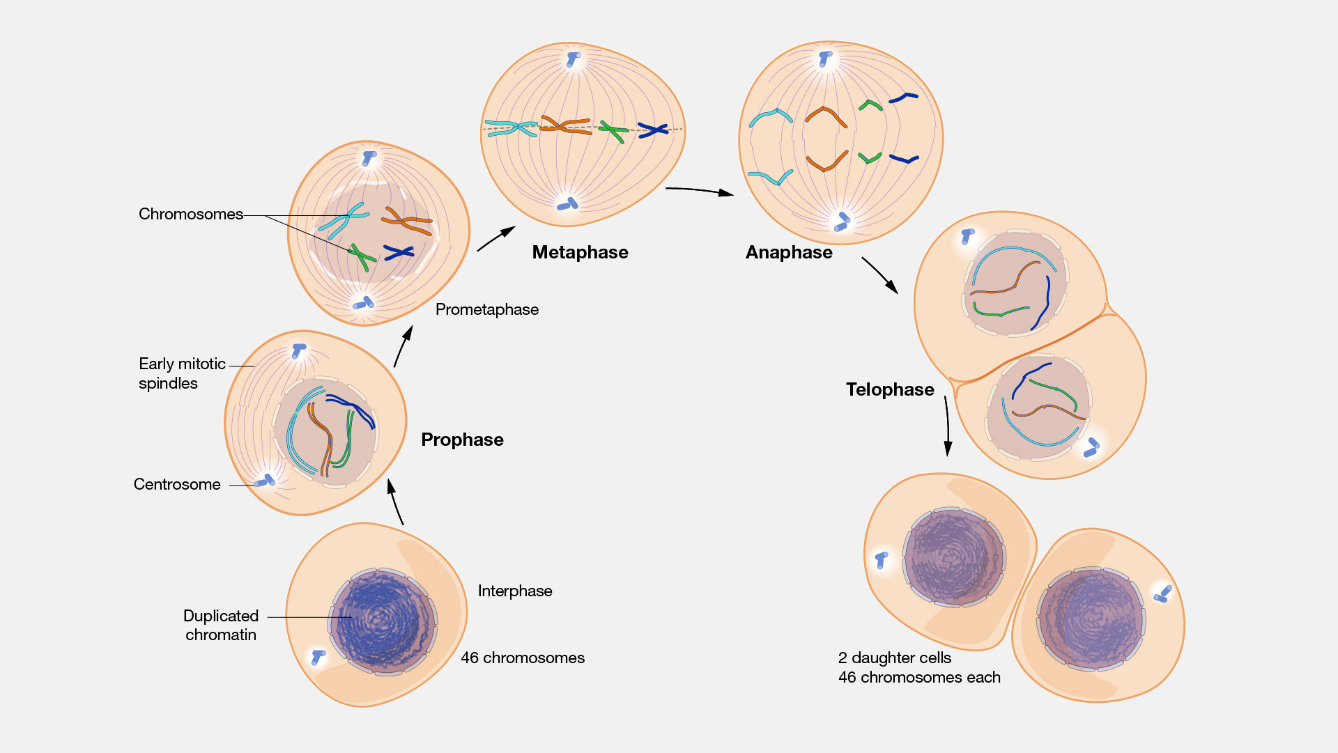
Cell division, specifically mitosis, is a bit like a production line for new cells, and it moves through several distinct periods. Each one of these periods is a "phase," a specific point in the cell's cycle where certain things happen, you know, a particular stage in a process. It’s not just one big event; it’s a series of changes, each with its own look and purpose. When we draw these phases of mitosis on the whiteboard, it helps to show how the cell's internal parts, especially its genetic material, get ready for and then actually divide. It’s like watching a time-lapse video of a plant growing, where you see each step clearly, rather than just the final, fully grown plant. This kind of sequential change, where one appearance gives way to another, is pretty much what we mean by a phase in this context, really.
The whole point of mitosis is to make sure that when one cell splits, the two new cells get an exact, complete set of genetic instructions. This means the cell has to be very careful about how it organizes and then separates its chromosomes, which are like the instruction manuals for the cell. So, you know, these phases are the cell's way of keeping everything tidy and making sure no instruction manuals get lost or messed up during the copying process. It’s a very organized series of appearances, one after the other, that the cell manifests to our eye or mind as it gets ready to duplicate itself. We break it down into these steps to make it easier to follow, like learning a dance step by step, actually.
Prophase on the Whiteboard - How Does It Start?
Prophase is the first major period we look at when we consider the phases of mitosis on the whiteboard. In this particular stage, the cell begins to get its act together, so to speak. The genetic material, which usually looks like a messy tangle of spaghetti inside the cell's central control area, starts to coil up and become visible as distinct, rod-like structures. These structures are the chromosomes, and they’ve already made copies of themselves, so each one is made of two identical halves, joined together. You might draw them on the whiteboard as X-shapes, showing they're doubled up and ready for separation. It’s a bit like taking a pile of loose threads and winding them onto spools, making them much easier to manage later on, you know, a clear appearance of preparation.
- Coach Birkin Dupe
- Kevin Gates Forehead
- Chinese Paratroopers Land In Florida
- Diddy Carl Wilson
- Really Hairy Lesbians
Also during this particular period, the structure that holds the genetic material, the nucleus, starts to break down. It kind of disappears, making way for the next steps. At the same time, tiny structures called centrosomes, which are like the cell's organizing centers, begin to move to opposite ends of the cell. From these centrosomes, long protein fibers, a bit like tiny ropes, start to stretch out across the cell. When you sketch this on the whiteboard, you show the chromosomes becoming clear, the nucleus fading, and these organizing centers beginning their journey to the cell's poles. This distinct period sets the stage for everything that comes next, a very clear step in the process, really.
Metaphase - Lining Things Up on the Whiteboard
Following prophase, we enter the next particular period, which we call metaphase, one of the key phases of mitosis on the whiteboard. This is where things get incredibly organized inside the cell. Those tiny ropes, or spindle fibers, that started to form in prophase, now attach to each of the doubled chromosomes. And this is the really neat part: all the chromosomes line up right in the middle of the cell, forming a straight line, almost like soldiers standing at attention. You would draw this on your whiteboard as a perfectly straight row of those X-shaped chromosomes, right in the center of your cell circle. It’s a very specific appearance, a defining characteristic of this stage.
This alignment is super important, you know, because it makes sure that when the cell eventually pulls these chromosomes apart, each new cell gets exactly one copy of each. If they weren't lined up just so, there's a good chance some cells would end up with too many or too few genetic instructions, and that would be a problem. So, this stage is a period of very precise arrangement, a critical step in the entire sequence. When you are looking at the phases of mitosis on the whiteboard, metaphase is often the easiest one to spot because of this very neat, central line of chromosomes. It shows a particular stage in the gradual development of the cell division process, where everything is perfectly poised for the next big move.
Anaphase - Pulling Apart on the Whiteboard
After everything is perfectly lined up in metaphase, the cell moves into anaphase, another crucial period among the phases of mitosis on the whiteboard. This is the moment of separation. The tiny ropes, those spindle fibers we talked about, suddenly shorten. As they pull back, they literally split each doubled chromosome right down the middle. One half of each X-shaped chromosome, which we now call a chromatid, gets pulled to one end of the cell, while its identical twin gets pulled to the opposite end. On your whiteboard, you would show these separated chromosomes moving away from the center, heading towards the poles of the cell, kind of like two groups of people walking away from each other. It’s a very dynamic appearance, a clear sign of action.
This pulling apart is a very quick and decisive action. It ensures that each new cell will receive a complete and identical set of genetic information. It's a bit like dividing a deck of cards exactly in half, making sure each person gets an equal share. This particular stage is a defining moment in the entire cell division process, where the genetic material is finally distributed. So, when you are illustrating the phases of mitosis on the whiteboard, anaphase is characterized by these distinct, moving sets of genetic material, showing a clear period of physical separation within the cell, a very important step in the sequence of events.
Telophase - Two New Beginnings on the Whiteboard
Telophase marks the beginning of the end for the original cell, and the start for two new ones, making it a very interesting period among the phases of mitosis on the whiteboard. Once the separated genetic material has reached the opposite ends of the cell, the cell starts to reverse some of the changes that happened earlier. New nuclear coverings begin to form around each set of chromosomes at both ends of the cell. This means that two distinct nuclei, two separate control centers, start to appear within what is still one large cell. On your whiteboard, you would draw two new circles forming around the clusters of chromosomes at each pole, showing the re-establishment of these important structures. It’s a period of reassembly, a return to a more organized state for the genetic material.
At the same time, the chromosomes themselves begin to uncoil and become less distinct again, returning to their more relaxed, spaghetti-like state. The spindle fibers, those tiny ropes, also start to disappear. This particular stage is all about setting up the conditions for two individual cells to form. It’s a bit like packing up after a big event, getting everything ready for the next day. So, when you are showing the phases of mitosis on the whiteboard, telophase represents the moment just before the final split, where you can clearly see two distinct sets of genetic material, each with its own newly formed covering, waiting for the cell to divide completely. It’s a significant stage in the gradual development of new cells.
Why Do We Look at Phases of Mitosis on the Whiteboard?
Looking at the phases of mitosis on the whiteboard is a really effective way to grasp this fundamental biological process. It’s a visual aid that helps break down something that happens at a microscopic level into understandable, step-by-step actions. Just as we might sketch out the different periods of a project or the various appearances of a changing situation, drawing cell division helps us see the distinct stages. It allows us to point to specific parts of the process and explain what's happening, rather than just talking about it in abstract terms. This kind of visual representation makes it much easier for our minds to connect with the concept, you know, to really see the progression.
A whiteboard lets us highlight the key changes in each period. We can draw the chromosomes changing their shape, moving, and then separating. We can show the nucleus disappearing and reappearing. This visual story helps to solidify the sequence of events in our memory. It’s a bit like using a map to understand a journey, where each phase is a particular landmark or section of the road. It also allows for interaction; you can draw, erase, and redraw, helping to reinforce the learning. So, in some respects, it's about making an invisible, continuous process into a series of clear, discrete moments that we can better comprehend. It’s a way of making the complex appear simpler, actually.
Cytokinesis - The Final Split After the Phases of Mitosis on the Whiteboard
While often discussed alongside the main phases of mitosis on the whiteboard, cytokinesis is technically the final step that completes the cell division process. After telophase, where the two new nuclei have formed at opposite ends of the cell, cytokinesis is the physical splitting of the cell's main body, its cytoplasm. This is where one big cell finally becomes two separate, individual cells. In animal cells, the cell membrane pinches inward, forming what looks like a furrow, eventually dividing the cell into two. In plant cells, a new cell wall forms down the middle, creating a barrier between the two new cells. You would draw this on your whiteboard as the original cell literally cleaving into two distinct entities, each with its own nucleus and a complete set of genetic instructions. It’s the very last appearance, the final act of separation.
This particular period is essential because without it, you'd just have one cell with two nuclei, which isn't the goal of cell division. Cytokinesis ensures that each new cell is a complete, independent unit, ready to carry out its own functions. It's the point where the "two new beginnings" from telophase truly become two separate, functional cells. So, when you are illustrating the phases of mitosis on the whiteboard, cytokinesis is the crucial final action that transforms one parent cell into two daughter cells, making it a very clear stage in the process of creating new life. It marks the end of one cycle and the start of two new ones, really.
What Happens If These Phases Go Wrong?
Thinking about the phases of mitosis on the whiteboard also brings up an important question: what if something goes amiss during these carefully orchestrated periods? Because each stage is so precise, any error in the sequence or in the actions of the cell's components can have significant consequences. For example, if the chromosomes don't line up correctly in metaphase, or if they don't separate evenly in anaphase, the resulting new cells might end up with too many or too few chromosomes. This is a bit like getting an incomplete or incorrect set of instructions for building something, you know, it just won't work right.
Such errors in cell division can lead to various issues for a living thing. For instance, in humans, an incorrect number of chromosomes in a cell can contribute to developmental problems or certain conditions. The cell has internal checks and balances, kind of like quality control steps, to try and catch these mistakes. But sometimes, these checks don't work perfectly. So, the study of these distinct periods of change, these phases, helps us understand not just how cells normally grow and repair, but also what happens when that process doesn't follow its usual path, which is pretty important, actually.
The journey of a cell dividing, as seen through the distinct phases of mitosis on the whiteboard, gives us a clear picture of how one cell becomes two. From the initial coiling of genetic material in prophase to its precise alignment in metaphase, then the crucial separation in anaphase, and finally the formation of new nuclei in telophase, each period is a vital step. Cytokinesis then completes the physical split, ensuring two fully separate cells. This step-by-step view, visually represented, helps us grasp the fundamental process of growth and repair in living things.

/stages-of-mitosis-373534-V5-5b84992cc9e77c00574f03d3.png)
Detail Author:
- Name : Maritza Lang
- Username : karli95
- Email : camille23@yahoo.com
- Birthdate : 2004-12-23
- Address : 3129 Bradtke Mountain Marshallfurt, IL 44909-4847
- Phone : 616-512-7969
- Company : Parker Inc
- Job : Petroleum Pump Operator
- Bio : Unde sed ipsa fugit perferendis delectus. Eius non et alias sit molestiae et et. Unde odio consequuntur consequatur qui sapiente perferendis ullam laboriosam.
Socials
linkedin:
- url : https://linkedin.com/in/crystelbarton
- username : crystelbarton
- bio : Ut ut doloremque tempora et.
- followers : 5138
- following : 737
facebook:
- url : https://facebook.com/crystel7263
- username : crystel7263
- bio : Et velit sed quisquam dolores est ut. Cum sint sit repudiandae veritatis.
- followers : 3099
- following : 97
